


Wound Healing Effects Of The Leaves Extract Of Aspilila Africana On Wistar Rats (Rattus norvegicus)
EA Osunwoke1*, OL Otakore1, SA Lelei2
1Department Of Human Anatomy, Faculty Of Basic Medical Sciences, University Of Port Harcourt, Nigeria
2Department Of Human Physiology, Niger Delta University, Nigeria
*Author for correspondence
Email : aeosunwoke@yahoo.com
Phone no: +2348055160338.
ABSTRACT
Background: The leaves extract of Aspilia africana was studied for its potency on experimentally induced wound in rats. Aim : To investigate the effectiveness of Aspilia africana as a wound healing agent. Methods: Wounds were inflicted on wistar rats using excision model. 20 male wistar rats (two groups of 10 control and 10 experimental rats) were used for this study. The wounds were processed for histological analysis. Healing was assessed by wound contraction rate, complete day of wound closure and histological analysis of excised tissue. Result: The wound contraction rates of experimental group on day 6 and 9 were statistically significant (P< 0.05). The mean wound closure for the experimental group was 12.6±1.17cm while that of the control group was 15.0±1.86cm. Concentration of neutrophils and macrophages were intense in experimental group than the control group in tissue samples excised on day 6. Also, the experimental group exhibited more intense concentration of neutrophils, macrophages, fibroblasts and capillaries on day 9 than the control group. It was observed that shorter period of total wound closure and increased inflammatory response corroborate the use of Aspilia africana in the topical management of wound area with time. Conclusion: This study has indicated that extracts of leaves of Aspilia africana have good potentials for use in wound care and further provide a rationale for the use of the leaves of this plant in wound management.
Key words: Aspilia africana, Wound Healing, Wistar rats
INTRODUCTION
The mechanism of tissue healing is a complex biological process that involves a perfect and coordinated cascade of cellular and molecular events promoting tissue reconstitution [1]. The use of plants for traditional and local medical purposes is an established fact. Aspilia africana is one of the many indigenous plants used in folk medicine to curb certain illness. The plant is a perennial herb varying in height from 60cm to about 1.5m depending on rainfall [2]. The extract of leaves of the plant has been implicated in the treatment of rheumatic pain [3] anticoagulating activities [4] anti-malaria [P. falaciparium] infection [5] and anti-inflammatory effect [6]. Aspilia africana was classified as a low toxicity drug after acute toxicity was carried out by administering oral doses of (2, 4, 8, 12 and16) g/kg body weight on wistar rats[7]. The anti-microbial properties of the leaf extract was demonstrated when petroleum ether, choloform and methanol extract was used in-vivo to treat bacterial (clinical strains of Staphylococcus and bacillus and clinical strains of Escherichia coli, Pseudomonas aeruginosa, Candida albicans and Aspergillus flavus) infections. The petroleum ether extract was most effective and had wide spectrum activities against all tested micro-organisms followed by the chloroform extract[8]. The leaf extract of Aspilia africana posses anti-inflammatory activity in acute and certain chronic inflammation, which was indicated from the inhibition of prostagladins synthesis, inhibition of increased vascular permeability, inhibition of neutrophil migration into inflamed tissues, and stimulation of lymphocyte accumulation which may enhance tissue repair and healing [6]. The terpenoids present in the leaves may account for the anti-inflammatory activity [9]. The anti-ulcer activity of aqueous leaf extract of Aspilia africana was investigated in ethanol, indomethacin and aspirin induced ulcer in wistar rats. The result showed significant reduction in the ulceration which was dose-dependent. The percentage ulcer inhibition of the extract at 100mg/kg for ethanol was 73.0%; 60.9% for indomethacin and 87.6% for aspirin [10]. Wound healing activity of gel formulations of methanol extract of leaves of Aspilia africana was studied for its potency on experimentally-induced wound in rats. Results showed that there was progressive decrease in wound area with time and a 100% wound closure was observed by the 17th post wound day in both gel formulations of extract and the standard which indicated the wound healing abilities of extract formulated in gel forms [11]. Some researchers have investigated the chemical constituents of Aspilia africana and documented it as follows; alkoloids (6.35±0.841%), tannins (0.1888 ±0.035%), flavonoids (2.006 ±0.11%), and phenols (0.109 ±15%). The concentration per 100g of vitamins in the plant are follows ascorbic acid (11.00± 0.15mg), niacin (3.045 ±0.081mg), thiamine (1.940 ±0.025mg), riboflavin (0.135 ± 0.100mg). The mineral elements determined per 100g, calcium (246±4.345mg), nitrogen (213 ±6.600mg), and phosphorus (25.00±0.500mg), zinc (36.675 ±0.54), nickel (10.975 ±1.330), selenium (Se) (10.875 ±1.880), Boron (B) (9.675 ±2.045) and lead (Pb) (7.425±1.050). They concluded that from the results it is possible that Aspilia africana posses medicinal potentials as claimed by traditional medicine practitioners [2,12].
The aim of this study is to investigate the effectiveness of Aspilia africana as a wound healing agent.
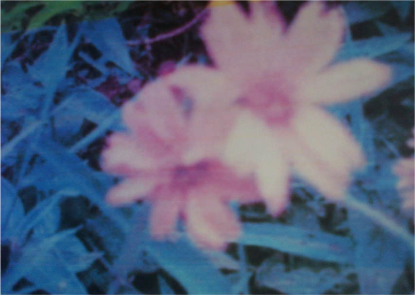
Photograph showing leaves of Aspilia Africana.
MATERIALS AND METHOD
Plant collection and identification
Fresh leaves of Aspilia africana were collected within the University of Port Harcourt. Plant was identified and authenticated at the Plant Science and Biotechnology Department of the same University.
Preparation of extract
Harvested fresh leaves were dried and pulverized to fine powder. 200 g of dried material was macerated in 3 liters of distilled water for 48hrs at 100 °C. Extract was sieved and filtered using a filter paper and then stored in a labeled air tight container.
Animal grouping and administration of extract
A total of 20 male adult wistar rats of average weight 200-220gms with absence of pre-existing skin lesion were used for the study, female rats were excluded because of hormonal variations. The rats were divided into 2 groups; Control group A and experimental group B having 10 rats each. They were kept for 2weeks to acclimatize, fed with standard rat cubes and water ad libitum. Using insulin syringe, 0.2mg of mixture of ketamine and diazepam each of equal quantity was used to induce unconsciousness. Wound incision was done on the right dorso-lateral flank of the rat using a sterile blade, taking care the panniculus carnosus because of its role facilitating wound contracture in rats [13,14].
A transparent paper of 2cmx2cm template was made and the outline on the skin traced with a permanent marker. Wounds of the control group were treated with normal saline solution while that of experimental group were treated with Aspilia africana extract. Wounds were then covered with sterilized gauze to avoid infection. Dressing of the wounds were done on a 3day interval (both control and experimental group). On day 9, five rats were randomly selected from each group for biopsy to obtain granulation tissues for histopathological and histomorphometric analysis. When the wounds finally healed the end scar tissue was also harvested for analysis.
The excised tissue was preserved using 10% formal saline and routine tissue processing was carried out and paraffin wax sections were prepared. Wound Size Measurement was done by tracing the wound with a marker on a sterile transparency.
RESULTS
The result of this study is as presented in the Table 1 below. Table showing the percentage mean wound contraction on day 6, 9 and the day of complete re-epithelialization. The wound was completely healed on day 18 for the control group and on day 12 for the experimental group.
Tables 1: Table showing percentage (%) mean wound contraction for control and experimental at day 6 and 9 and mean for complete re-epithelialization.
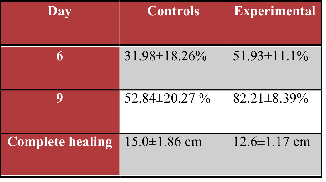
Plate1: Plate showing photomicrograph of control animals on day 9; H&E x200. The section of the skin shows moderate fibrosis, marked with infiltration of neutrophils and macrophages.
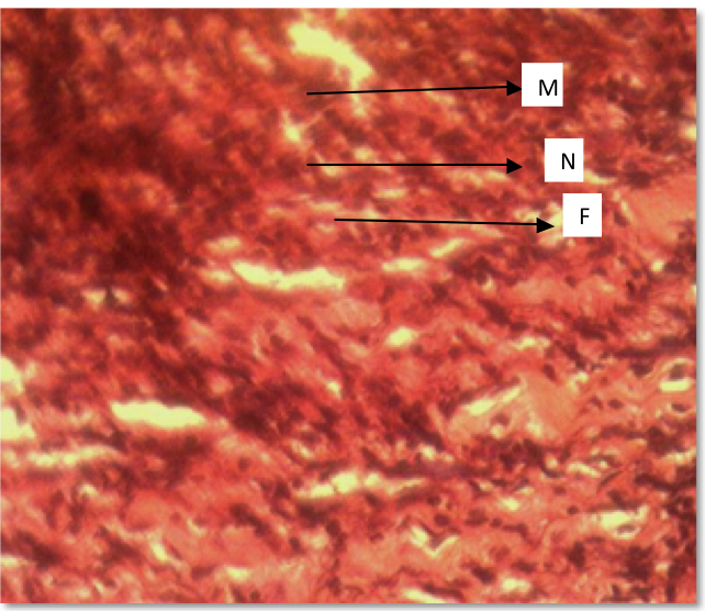
Plate1: Photomicrograph of control day 9: H&E x200; (F) = Fibrosis, (M) = Macrophage (N) = Neutrophil
Plate2: Plate showing photomicrograph of control animal end scar; H&E x200. The section of the scar tissue shows an atrophic epidermis with decreased keratinization, a markedly fibrous dermis and moderate infiltration of inflammatory cells.
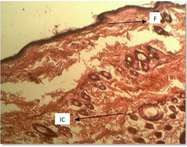
Plate2: Photomicrograph Of Control End scar: H&E X200; (IC) = Inflammatory cells, (F) = Fibrosis
Plate3: Plate showing photomicrograph of experimental animals on day 9; H&E x200. This section of the skin shows a healing ulcer with marked infiltration of inflammatory cells, mild fibrosis and neovascularization.
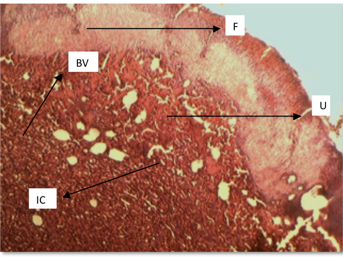
Plate3: Photomicrograph Of Experimental Day 9: H&E X200; (U) = Ulcer, (IC) = Inflammatory cells, F= Fibrosis, BV= Blood vessels.
Plate 4: Plate showing photomicrograph of experimental animal end scar: H&E x200. This section of the scar tissue shows hyperkeratosis, hypergranulosis and acanthosis. Numerous hair follicles and some sebaceous glands were observed.
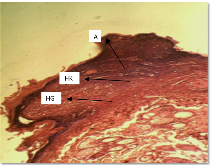
Plate 4: Photomicrograph Of Experimental End scar: H&E X200; (HK) = Hyperkeratosis, (A) = Acanthosis, HG= Hypergranulosis.
DISCUSSION
There was a significant percentage wound contraction, fibroblast and blood vessels count in the microscopic appearance of granulations and end scar tissue of the wounds dressed with the extracts of Aspilia africana leaf extract. High intensity of inflammatory, infiltration of neutrophils, macrophages and numerous blood vessels were observed in the experimental group and less in the control group. Significant wound contraction and complete re-epithelialization thus indicating that the leaf extract possess wound healing properties which agrees with the study of some researchers [8]. The observed decrease in the wound area on the application of the extract of Aspilia Africana indicated that the plant extract possesses wound healing properties [6]. Aspilia Africana has been reported to possess antimicrobial and haemostatic activities which are necessary in wound healing process. Since wound provide an environment for microbial growth, the antimicrobial activity of the extract may partly contribute to the wound healing effect by eliminating infections thus allowing the natural tissue repair processes to start [15],
The leaf extract of Aspilia africana promoted blood clothing in a short time which is an important index of haemostatic activity as supported by the work of another researcher who stated that the leaf extracts of Aspilia africana may have facilitated the haemostatic process to effect wound healing, promoted angiogenesis and neovascularization which is a crucial step in wound healing [16]. This is in line with the reports that high density capillary also suggests increased angiogenesis [17]. Wounds treated with the extracts of Aspilia africana showed the presence of fibrosis in the dermis and hyperkeratosis (increased thickness of the stratum corneum) which is as a result of collagen formation and angiogenesis that is necessary for wound healing [18,19]. At the end of the experiment, there were no hypertrophic scars in the wounds generated.
CONCLUSION:
This research has shown that the aqueous extracts of the leave of Aspilia africana promotes wound healing activity through increased inflammatory response and neovascularization. It will be worthwhile to further carry out a concentration-based study to evaluate the effects of the extracts of Aspilia africana.
Further studies with isolated constituents are required to understand the complete mechanism of wound healing activity of Aspilia africana.
ACKNOWLEDGEMENT
I wish to acknowledge all those who contributed in no little way to the success of this study.
REFERENCES
| 1. Singer AJ, Clark R: Cutaneous wound healing. N Engl J Med 1999, 341:738-746. http://dx.doi.org/10.1056/NEJM199909023411006 PMid:10471461 |
||||
| 2. Abu T, Onuoha E: The chemical constituents of the leaf of Aspilia africana as a scientific backing to its tradomedical potentials. Agricultural Journal 2011, 6:28-30. http://dx.doi.org/10.3923/aj.2011.28.30 |
||||
| 3. Oliver-Bever B: Medicinal Plants in Nigeria: Being a Course of Four Lectures Delivered in April 1959 in the Pharmacy Department of the Nigerian College of Arts, Science and Technology, Ibadan: Published as a private ed. by the Nigerian College of Arts, Science and Technology; 1960. | ||||
| 4. Hanna MM, Niemetz J: Studies on the anticoagulant action of Aspilia africana. Thrombosis research 1987, 47:401-407. http://dx.doi.org/10.1016/0049-3848(87)90455-5 |
||||
| 5. Okokon J, Nwidu L, Essiet G: Evaluation of in-vivo antiplasmodial activity of Aspilia africana. Inter. J. Pharm 2006, 2:348-351. http://dx.doi.org/10.3923/ijp.2006.348.351 |
||||
| 6. Okoli C, Akah P, Okoli A: Potentials of leaves of Aspilia africana (Compositae) in wound care: an experimental evaluation. BMC Complementary and Alternative Medicine 2007, 7:24. http://dx.doi.org/10.1186/1472-6882-7-24 PMid:17623087 PMCid:PMC1940021 |
||||
| 7. Taziebou LC, Etoa F, Nkegoum B, Pieme C, Dzeufiet D: Acute and subacute toxicity of Aspilia africana leaves. African Journal of Traditional, Complementary and Alternative Medicines 2008, 4:127-134. http://dx.doi.org/10.4314/ajtcam.v4i2.31203 |
||||
| 8. Adeniyi B, Odufowora R: In-Vitro Antimicrobial Properties Of Aspilla Africana (Compositae). 2000. | ||||
| 9. Okoli C, Akah P, Nwafor S, Anisiobi A, Ibegbunam I, Erojikwe O: Anti-inflammatory activity of hexane leaf extract of Aspilia africana CD Adams. Journal of ethnopharmacology 2007, 109:219-225. http://dx.doi.org/10.1016/j.jep.2006.07.037 PMid:16950582 |
||||
| 10. Ubaka M, Ukwe V, Okoye C, Adibe O: Investigation into the anti-ulcer activity of the aqueous leaf extract of Aspilia africana CD Adams. Asian Journal of medical sciences 2010, 2:40-43. | ||||
| 11. Attama A, Uzor P, Nnadi C, Okafor C: Evaluation of the wound healing activity of gel formulations of leaf extract of Aspila africana Fam. Compositae. Journal of Chemical and Pharmaceutical Research 2011, 3:718-724. | ||||
| 12. Oko O, Agiang E, Osim E, Asuquo O: Toxicological Evaluation of Aspilia Africana Leaf Extracts in Mice. American Journal of Pharmacology and Toxicology 2011, 6:96-101. http://dx.doi.org/10.3844/ajptsp.2011.96.101 |
||||
| 13. Billingham R, Russell P: Studies on wound healing, with special reference to the phenomenon of contracture in experimental wounds in rabbits' skin. Annals of surgery 1956, 144:961. http://dx.doi.org/10.1097/00000658-195612000-00005 PMid:13373285 |
||||
| 14. Badoe E, Archampong E, Jaja M: Principles and practice of surgery. Liver and Biliary System, Ghana Publishing Corporation, Tema 1994:657-688. | ||||
| 15. Achonye E: A pharmacological investigation of the haemostatic action of pressed leaf extract of Aspilia latifolia (Compositae). B. Pharm thesis University of Nigeria. Pharmacology & Toxicology Department 1976. | ||||
| 16. O'Reilly R: Drugs used in disorders of coagulation. Basic and Clinical Pharmacology 1998, 17:547-561. | ||||
| 17. Clark R: Cutaneous wound repairs. Physiology, Biochemistry and Molecular Biology of Skin. Goldsmith LA. Edited by: Oxford University Press, New York; 1991. | ||||
| 18. Trabucchi E, Preis BF, Baratti C, Montorsi W: Topical treatment of experimental skin lesions in rats: macroscopic, microscopic and scanning electron-microscopic evaluation of the healing process. International journal of tissue reactions 1986, 8:533. PMid:2432029 |
||||
| 19. Shukla A, Rasik AM, Dhawan BN: Asiaticosideâ€induced elevation of antioxidant levels in healing wounds. Phytotherapy Research 1999, 13:50-54. http://dx.doi.org/10.1002/(SICI)1099-1573(199902)13:1<50::AID-PTR368>3.0.CO;2-V |
||||
![]()

Dr E.A Osunwoke holds a PhD in human Anatomy. He is a lecturer in the Department of Human Anatomy, Faculty of Basic Medical Sciences, University of Port Harcourt, Rivers state, Nigeria. He has many research articles published in both local and international journals.